Imaging
15 min read
By BioBuilt Editorial

Imaging
15 min read
By BioBuilt Editorial
For most of human history, the only way to understand what was happening inside the body was to open it. A physician facing a patient with unexplained pain had little to work with beyond a pulse, a fever, the color of the skin, and educated guesswork. When those clues weren't enough, and they often weren't, the only option left was exploratory surgery, cutting a patient open not because the surgeon already knew what needed fixing, but specifically to find out, with no anesthesia to spare the patient and no antiseptics to guarantee they'd survive the exploration itself.
Physicians spent centuries developing clever workarounds for reading the body from the outside instead: tapping on the chest to listen for the dull thud of fluid where there should have been the hollow resonance of healthy lung, pressing an ear or a hollow wooden tube against the ribs to catch the rhythm of a heartbeat. Every one of these techniques was an attempt to guess at hidden anatomy through indirect clues, because there was, quite literally, no other way to know. Today, physicians can watch blood flow through arteries, map neural activity across the brain, detect tumors only millimeters wide, and observe individual molecules inside living tissue, all without making a single incision. None of that ability came from biology. It came from engineering.
Every medical imaging technology in use today is really an answer to the same underlying question, asked in a different way: how do you see something that light can't reach? The skin, muscle, and bone that make the human body opaque to our eyes don't stop other kinds of signals, radiation, sound, magnetism, chemistry, if you know how to send them in and read what comes back out. The history of medical imaging isn't a story about biology becoming better understood. It's a story about engineers inventing new definitions of what "seeing" can mean.
In 1895, the German physicist Wilhelm Röntgen noticed that a mysterious form of radiation could pass through solid objects and expose photographic film on the other side, and that different materials blocked it by different amounts. He'd discovered X-rays, and within weeks, doctors were using them to look at broken bones. But the real engineering insight wasn't the discovery of a new kind of radiation. It was the discovery that the body itself is not uniformly transparent to it.
Dense tissue, like bone, absorbs X-rays strongly, so little radiation passes through to expose the film behind it, leaving that region pale on the image. Soft tissue absorbs less, letting more radiation through. Air, as in the lungs, absorbs almost none at all, so that region appears nearly black. What you're looking at in an X-ray isn't really a picture of anatomy. It's a map of attenuation, a record of how much of the beam each part of the body managed to stop. Learning to read that map, and later to engineer film and detectors sensitive enough to capture subtler differences, turned a strange laboratory curiosity into medicine's first real window into the living body.
That single insight, that different tissues interact with a signal differently, and that the differences themselves can be turned into an image, became the founding principle of an entire field. Every imaging technology that followed X-rays, in some form, is a variation on it.

A single X-ray has an obvious limitation: it flattens a three-dimensional body into a two-dimensional shadow. A tumor sitting behind a rib might be invisible, hidden in the overlap of everything the beam passed through on its way to the film. Solving that problem took a different kind of insight, one that came less from physics than from mathematics.
If one X-ray image collapses the body from every angle into a single flat view, what happens if you take hundreds of X-ray images, from hundreds of different angles around the body, and feed them all into an algorithm designed to reconstruct the three-dimensional structure that could have produced exactly that set of two-dimensional shadows? That's computed tomography, or CT, and it's a genuine engineering triumph disguised as a medical device. The scanner itself, rotating an X-ray source and detector around the patient, is almost the easy part. The hard part is the reconstruction: an inverse problem, working backward from a large set of measurements to infer the hidden three-dimensional structure that generated them, solved computationally rather than optically.
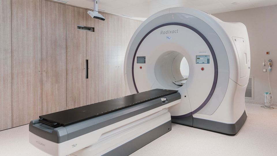
That reconstruction problem hasn't stood still. Engineers have since pushed CT-style reconstruction beyond static diagnostic scans and into the operating room itself, building compact cone-beam CT systems that can generate 3D images in real time to guide a surgeon's instruments or track a needle as it moves through soft tissue.
It's worth pausing on how CT was actually invented, because the story is a nice illustration of how unglamorous engineering breakthroughs often are. Godfrey Hounsfield, an electrical engineer at the British company EMI, worked out the reconstruction algorithm and built the first working scanner in the late 1960s, work funded in part by profits EMI had earned from its recording artists, including the Beatles. In his own account, written years later for the Nobel committee, Hounsfield described the idea starting small: "one of the suggestions I put forward was connected with automatic pattern recognition," an idea that eventually grew into the EMI scanner. Around the same time, and entirely independently, the South African-born physicist Allan Cormack had already worked out much of the underlying mathematics for reconstructing a cross-sectional image from X-rays taken at many angles, without ever building a machine to test it. Neither man knew of the other's work until years later. In 1979, the two shared the Nobel Prize in Physiology or Medicine, one of the rare cases of that prize going to what is, at its core, a piece of applied mathematics and electrical engineering. The core mathematical trick, hundreds of 2D projections turned into one 3D volume, is the same one that made CT possible in the first place. What's changed is the ambition: instead of imaging a patient before treatment, engineers are now building systems that can image a patient during it.
X-ray and CT both work by sending some form of radiation through the body and measuring what gets absorbed. Ultrasound throws that whole approach out. Instead of light or radiation, it uses sound, and instead of measuring absorption, it measures reflection.
The technology depends on a property of certain materials called the piezoelectric effect: apply an electrical voltage to a piezoelectric crystal, and it physically deforms; squeeze or vibrate it, and it generates a voltage in return. An ultrasound probe uses this effect twice in rapid succession, first to convert an electrical pulse into a burst of high-frequency sound, and then, a fraction of a second later, to convert the returning echoes back into an electrical signal. Different tissues reflect sound differently, and the amount of time an echo takes to return reveals exactly how deep the reflecting surface was, since the speed of sound in tissue is well known. Send out a pulse, time the echoes, and you can build a live, moving picture of what's underneath the skin, in real time, with no radiation exposure at all.
The engineering challenge that came after the basic idea worked was steering and focusing the sound itself. Beamforming, the technique of firing an array of many small piezoelectric elements with precisely timed delays, allows engineers to focus and sweep an ultrasound beam electronically, without moving a single physical part. That single innovation is why modern ultrasound machines can produce a smooth, live video image instead of a single static reading.
The technology has kept shrinking since. Traditional ultrasound probes rely on individual piezoelectric crystals, delicate components that are expensive to manufacture and easy to damage. In 2011, the entrepreneur Jonathan Rothberg founded Butterfly Network with a different approach: replacing those crystals with an array of thousands of microscopic vibrating membranes etched directly onto a semiconductor chip, the same manufacturing process used to make computer processors. The result, a handheld ultrasound probe that plugs into a smartphone, uses software to make the single chip mimic several different kinds of ultrasound probes at once, work motivated in part by how difficult it had been for Rothberg's own family to access imaging during his daughter's medical care. "One of the ways you democratize things is by putting them on chips," he said of the approach. It's a good example of how much of the recent progress in medical imaging has come not from discovering a new physical signal, but from re-engineering the manufacturing of an old one.
Every technology discussed so far images anatomy, the physical shape and structure of tissue. Positron emission tomography, or PET, does something stranger: it images chemistry.
PET works by injecting a patient with a small amount of a molecule, most commonly a modified sugar called fluorodeoxyglucose, or FDG, that has been tagged with a radioactive atom. Because FDG resembles the glucose cells normally burn for energy, it gets taken up most eagerly by whichever cells are working hardest, and cancer cells, which tend to consume glucose at an unusually high rate, often light up brightly on a PET scan long before a tumor is large enough to see on a CT or MRI. As the radioactive tag decays, it emits a particle called a positron, which almost immediately collides with a nearby electron and annihilates, producing two photons that shoot off in exactly opposite directions. A ring of detectors surrounding the patient watches for these paired photons arriving at the same instant on opposite sides of the ring, and by triangulating thousands of these coincident pairs, the scanner reconstructs a three-dimensional map, not of anatomy, but of metabolic activity.
It's an entirely different kind of "seeing," one aimed at function rather than form. Engineered nanoparticles designed to bind specific disease-related molecules, then be detected through magnetic, optical, or radioactive signals, are one active path building on that same idea, allowing a broad panel of biomarkers to potentially be read out from a sample far smaller than conventional methods require. The underlying goal is the same one PET was built around: making a chemical signature, not just a shape, into something an engineer can capture and turn into an image.
PET itself came out of a specific engineering partnership. The chemist Michael Phelps and his postdoctoral researcher Edward Hoffman built the first working PET scanner in 1973 at Washington University in St. Louis, combining three separate insights: a ring of detectors that could catch pairs of photons arriving in coincidence, a mathematical algorithm for turning those detections into a three-dimensional image, and the realization that positron-emitting versions of common biological elements, oxygen, nitrogen, carbon, and fluorine, could be used to label real biological molecules and track their fate inside a living body. That last insight is what allowed FDG, a radioactively tagged sugar, to become PET's most widely used tracer. Phelps has described the underlying goal of the technology as studying disease biology inside what he calls "the ultimate laboratory, the living human body."
Magnetic resonance imaging is, by most measures, the strangest entry on this list, and also one of the most powerful. It produces detailed, high-contrast images of soft tissue with no radiation at all, using nothing but magnetic fields and radio waves.
The trick starts with hydrogen, which is abundant throughout the body in water and fat, and whose nucleus behaves like a tiny spinning magnet. Placed inside the powerful magnetic field of an MRI scanner, these countless hydrogen nuclei align themselves, mostly, along the direction of the field. A precisely tuned radio-frequency pulse then knocks them out of alignment, and as they relax back into place, they release a faint radio signal of their own. Different tissue types relax at different rates, a property engineers call T1 and T2 relaxation, and those differences in timing are what give MRI images their remarkable ability to distinguish between soft tissues that look nearly identical on a CT scan.
The genuinely difficult engineering problem in MRI isn't producing that signal, it's turning a chorus of billions of these faint radio echoes into a coherent picture. By cleverly varying the magnetic field across the body in a controlled way, engineers can encode spatial position directly into the frequency and phase of the returning signal, and then use a mathematical technique related to the Fourier transform to unscramble that encoded signal back into an image, pixel by pixel. It's a system built almost entirely out of applied physics and computation, with barely any resemblance to a camera at all, and yet the result is some of the most detailed pictures of the human body's soft tissue that exist.
The chemist Paul Lauterbur worked out the key trick in 1971, and by his own account, the idea arrived while he was eating a hamburger at a restaurant, turning over a colleague's recent finding that nuclear magnetic resonance signals differed between healthy and cancerous tissue. His insight was that adding a gradient, a magnetic field that varies slightly and predictably across space, made it possible to tell exactly where in the body a given radio signal was coming from. He was reportedly in such a hurry to get the idea down that he sketched his notes on a napkin before formalizing them in a notebook days later. The British physicist Peter Mansfield then developed the mathematics to process that signal quickly enough, and the imaging sequences, to turn the idea into something fast enough for real patients rather than hours-long laboratory experiments. Lauterbur later titled his Nobel lecture "All Science Is Interdisciplinary," a fitting label for a discovery that came from a chemist, refined by a physicist, and delivered to medicine. The two shared the 2003 Nobel Prize in Physiology or Medicine for the work, three decades after the original insight, roughly how long it took the engineering of gradients, magnets, and computation to catch up to the physics.
Every technology so far images the body at the scale of organs, tissues, or, at best, small structures within them. But some of the most important recent progress in medical imaging has gone the opposite direction, toward the scale of individual cells, and this is where engineering and biology start to blend almost completely.
Fluorescence microscopy is the foundation of this shift: attach a molecule that glows under a specific wavelength of light to a target of interest, whether a protein, a strand of DNA, or an entire cell type, and suddenly you can see exactly where that target is and how much of it is present, against a dark, otherwise invisible background. Confocal microscopy improved on ordinary fluorescence imaging by adding a small pinhole aperture that blocks out-of-focus light, allowing researchers to capture crisp optical "slices" through thick tissue rather than a single blurred composite. Multiphoton microscopy pushed further by using infrared light, which penetrates deeper into living tissue and causes far less photodamage, making it possible to watch cellular processes unfold in living animals over extended periods. Super-resolution microscopy, a genuine physics breakthrough recognized with the 2014 Nobel Prize in Chemistry, shared by Eric Betzig, Stefan Hell, and William Moerner, found clever ways around the diffraction limit, a fundamental physical barrier that once made it impossible to see structures smaller than roughly half the wavelength of light being used. And light-sheet microscopy solved a different problem entirely, speed and gentleness, by illuminating only a single thin plane of a sample at a time, which allows researchers to image entire living embryos or organoids in three dimensions without bathing the whole sample in damaging light.
What ties these techniques together is a trend that's reshaping the entire field: microscopy is no longer just about better optics. Increasingly, it's about pairing optical systems with intelligent computation, using algorithms not merely to clean up an image after it's captured, but as an integral part of how the image is formed in the first place. Rather than treating optics and software as two separate stages, one to capture an image and one to interpret it, researchers increasingly design the optical system and the algorithm together, aiming the combination at problems where a purely optical instrument would fall short: catching a growth too subtle for the naked eye during an endoscopic exam, for instance, or extracting information from light that a standard camera would simply discard. The camera and the algorithm, in this approach, aren't separate steps. They're one integrated instrument.
If the last century of medical imaging was about engineers inventing entirely new signals to send into the body, X-rays, sound, magnetism, radioactive tracers, the next chapter looks increasingly like it will be about combining those signals, and increasingly handing part of the "seeing" over to computation itself.
Photoacoustic imaging is one of the clearest examples: shine a pulse of laser light into tissue, and different molecules will absorb it and heat up by a tiny, safe amount, causing a rapid, localized expansion that generates a faint sound wave. Detect that sound wave with an ultrasound-style sensor, and you get an image with the rich chemical contrast of optical imaging, but the depth and resolution of ultrasound, a hybrid technique that simply didn't exist as an option before engineers realized light and sound could be combined this way.
Much of the technique's development traces back to the work of Lihong Wang, a biomedical engineer now at Caltech, whose lab has spent more than two decades building out the method, from early 3D photoacoustic microscopy to functional photoacoustic tomography capable of imaging blood oxygenation and activity across the entire human brain. Wang has explained the core limitation photoacoustic imaging was built to solve simply: light scatters so badly inside tissue that "this is why we can't even see our own bone in the hands," even though the light is right there, just past the skin. His group has also built cameras that push the very idea of "seeing" to its physical limit: a technique called compressed ultrafast photography can capture roughly ten trillion frames per second, fast enough to record the passage of a single pulse of light itself. It's a useful reminder that "imaging" isn't a fixed target engineers eventually finish. Every gain in speed or contrast tends to reveal a new problem worth solving.
Computational imaging, more broadly, treats the optical or electronic hardware and the reconstruction algorithm as a single system to be designed together, rather than treating better hardware and smarter software as separate problems to solve independently. AI-based reconstruction is one of the most active fronts here, using machine learning models trained to recover full, high-quality images from far less raw data than traditional reconstruction methods require, an approach already being used to speed up MRI scans, sharpen low-dose CT images, and even correct for tissue that shifts or deforms during a procedure by registering preoperative scans against live imaging in the operating room.
Molecular imaging continues to push toward earlier and more specific disease detection, using engineered nanoparticles and novel sensing chemistries to catch a cancer's chemical signature before it ever grows large enough to be seen anatomically. Point-of-care and wearable imaging aims to take technologies that once required a hospital-sized machine and shrink them down to something portable and affordable enough to reach clinics with little infrastructure at all, handheld and low-cost devices designed to bring a meaningful slice of diagnostic capability to places a full imaging suite will never reach. Intraoperative imaging is moving imaging out of the diagnostic suite entirely and into the middle of surgery itself, giving surgeons a real-time, updating view of anatomy that shifts and deforms as they operate, guided in some cases by robotic systems that combine imaging with autonomous or semi-autonomous instrument control. And engineers working in soft robotics are beginning to build small, flexible, sensor-equipped devices designed to navigate through the body's own pathways, hinting at a future where the sensing and imaging systems that observe a patient might not stay outside the body at all.
None of these future directions replaces what came before. Photoacoustic imaging doesn't retire ultrasound, and AI reconstruction doesn't retire MRI physics. Each new technique is another answer, engineered rather than discovered, to the same question that's driven the whole field since Röntgen's photographic plates first exposed a shadow of the bones inside a hand: how do you see something that light can't reach?
It's tempting to think of medical imaging as a supporting technology, the thing that happens before the real medicine, the diagnosis or the surgery or the treatment, begins. But that framing undersells what's actually been built. Every one of these technologies expanded what a physician is capable of measuring in a living human being, and every expansion opened up diseases that couldn't be caught early, structures that couldn't be operated on safely, or biological processes that couldn't be studied at all until someone figured out how to make them visible.
Biomedical engineering is often described, understandably, as the field that builds new therapies: artificial organs, gene-editing tools, engineered cells. But making the invisible visible is just as fundamental a form of engineering, and arguably a prerequisite for everything else. You cannot treat what you cannot see. The story of medical imaging is the story of engineers steadily refusing to accept that limitation, one new signal, one new algorithm, one new way of looking at a time.
monthly newsletter
Get one monthly simplified summary of the healthcare headlines, research findings, and biotech developments worth understanding.